How the Eye Works
Vision is a complex sense composed of many elements. The human eye, elegant in its detail and design, represents a gateway to the process we call vision. The eyeball, or globe, is spherical in shape and about 1 inch across. It houses many structures that work together to facilitate sight.
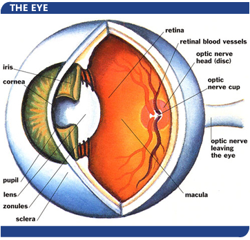 The human eye is comprised of layers and internal structures, each of which performs distinct functions. The outside layer of the eye is comprised largely of a tough, white, protective tissue called the sclera. The sclera helps maintain the shape of the eyeball. At the front of the eye is an equally tough but clear structure called the cornea, which is responsible for letting light into the eye and bending light. The human eye is comprised of layers and internal structures, each of which performs distinct functions. The outside layer of the eye is comprised largely of a tough, white, protective tissue called the sclera. The sclera helps maintain the shape of the eyeball. At the front of the eye is an equally tough but clear structure called the cornea, which is responsible for letting light into the eye and bending light.
Going from outside to inside, the next layer of the eye is the choroid, which carries the blood supply necessary to nourish the eye's internal structures. Finally, there is the layer called the retina, lining the inside of the eye, which is sensitive to light and receives stimulation to its specialized cells.
The eye has a number of protective features. The eyelids, eyelashes and eyebrows are all designed to protect the eye from dirt and dust that might enter it and cause damage. The globe sits inside the orbital cavity, a bony pocket lined with fatty tissue as a cushion. Together these provide additional protection against injury. Six muscles attach at various points to the sclera and enable the globe to move in many directions inside the orbit.
In order for vision to take place, a succession of processes must occur involving the structures within the eye and the brain:
The first part of this chain is that light rays must travel through the eye to ultimately focus on the retina. There are a number of structures involved in the bending or refracting of light so that it focuses properly. Light first passes through the clear cornea at the front of the eye, and then through a watery substance called the aqueous humor which fills the small chambers located behind the cornea. As light continues on its pathway it passes through the pupil, a round opening in the center of the iris. The iris is the part of the eye that gives the eye its color. It also is made up of specialized muscles that are able to change the size of the pupil from very small (about 2 mm) to large (about 8 mm), regulating the light that is entering.
The next structure light will penetrate is the lens, another clear, layered structure shaped like a large lentil (about 10 mm in diameter) that is attached to muscles which contract or relax to change the shape of the lens. The changing lens shape helps light to be focused in response to the need for clarity. (The loss of this focusing ability as humans age -- a natural occurrence -- is the reason that many adults over 40 years old need reading glasses.) Once through the pupil and lens, the light then passes through the larger posterior (back) portion of the eye that is filled with a clear, jelly-like substance called the vitreous humor. From there, the light will come to the retina, where the rod cells and cone cells will be stimulated to set off a chain of split-second chemical reactions converting light to electrical impulses. The cone cells (about 7 million in number) are located in greatest concentration in the small, central part of the retina called the macula. This area is responsible for producing sharp, detail vision and color vision. The rod cells (numbering about 100 million) are found in the peripheral retina, away from the macula. These cells provide vision in dim light.
Even if all of the structures of the eye work perfectly, what we know as vision cannot happen without the brain's interpretation of the electrical impulses sent by the retina. The optic nerve is the bundle of retinal fibers that exits the back of the eye and transports electrical impulses to the brain where they are interpreted in the primary visual cortex.
When all parts of the visual system are working, the eyes can move together, can adapt to light and dark, perceive color and accurately evaluate an object's location in space. They are sensitive to differences in contrast, and can also provide detail vision, which is measured as visual acuity. By convention, we know "normal" visual acuity to be reported as 20/20. As the bottom number of this expression gets higher, it tells us that the vision is poorer than "normal." For example, the start of the range known as "legal blindness" is represented by the visual acuity finding of 20/200. One way to understand the meaning of this finding is that the eye being tested sees at 20 feet what the "normal" eye would see at 200 feet. People whose vision is in the category of "legal blindness" may still be able to use vision to do some of the things they need to do.
All eyes are not the same, nor are they all perfect. Some eyeballs are too long or have too much focusing power, causing the person to be myopic (nearsighted). Others are too short or have too little focusing power, and the result is hyperopia (farsightedness). Some eyeballs may have uneven curvature, called astigmatism. Options for correcting these "mechanical" problems are standard eyeglasses, contact lenses or refractive surgery. Other problems may be caused by disease or injury, and are not correctable by conventional means. People whose vision is irreversibly impaired due to diseases such as macular degeneration, glaucoma, cataract, diabetic retinopathy and others can be helped by vision rehabilitation.
|